Lily flower bud, light micrograph

![]()
Wall Art and Photo Gifts from Science Photo Library
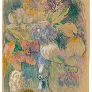
Lily flower bud, light micrograph
Lily flower bud. Light micrograph of a section through a young flower bud from a lily (Lilium sp.), showing pollen cells (circles) dividing within the anthers (inner ring). The outer floral envelope consists of an outer and inner whorl of three petals (perianth or tepals). Next are the male stamens arranged in two whorls of three, known as the androecium. The anthers, on the ends of the stamens, have four pollen sacs, within which the diploid sporophyte pollen grains are undergoing meiosis (cell division) to form the haploid gameteophyte. In the centre of the flower are the three fused female carpels, which form the gynaecium. Magnification: x3 when printed 10 centimetres wide
Science Photo Library features Science and Medical images including photos and illustrations
Media ID 6352581
© DR KEITH WHEELER/SCIENCE PHOTO LIBRARY
Androecium Anther Anthers Carpel Carpels Cell Biology Cell Division Cytological Cytology Diploid Dividing Fused Grains Histological Histology Lily Meiosis Meiotic Microscopy Monocot Monocots Monocotyledon Monocotyledons Outer Pollen Grain Pollen Sac Re Production Reproductive Structure Sacs Sporophyte Stain Stained Stamen Stamens Structural Structures Tepal Tepals Tissue Whorl Whorls Cells Light Micrograph Light Microscope Section Sectioned
EDITORS COMMENTS
This stunning print captures the intricate beauty of a lily flower bud at a microscopic level. The light micrograph reveals the fascinating structures and processes taking place within this young bud. At the center of the image, we can see three fused female carpels forming the gynaecium, which is surrounded by an outer and inner whorl of three petals known as perianth or tepals. Moving outward, we encounter two whorls of male stamens called androecium, with four pollen sacs on their ends. The highlight of this micrograph lies in the anthers where pollen cells are dividing through meiosis to form haploid gameteophytes. These tiny circles represent diploid sporophyte pollen grains undergoing cell division. The meticulous staining technique used for this image enhances our understanding of plant biology and botany. It allows us to appreciate not only the structural complexity but also provides insights into reproductive structures and processes in angiosperms like lilies. With a magnification factor of x3 when printed 10 centimeters wide, this photograph invites us to explore every detail meticulously captured by Science Photo Library's expert photographers. Whether you have an interest in cytology or simply admire nature's wonders, this print will undoubtedly captivate your imagination with its vibrant colors and intricate patterns.
MADE IN THE USA
Safe Shipping with 30 Day Money Back Guarantee
FREE PERSONALISATION*
We are proud to offer a range of customisation features including Personalised Captions, Color Filters and Picture Zoom Tools
SECURE PAYMENTS
We happily accept a wide range of payment options so you can pay for the things you need in the way that is most convenient for you
* Options may vary by product and licensing agreement. Zoomed Pictures can be adjusted in the Cart.