Home > Arts > Artists > J > Jacob Jacobs
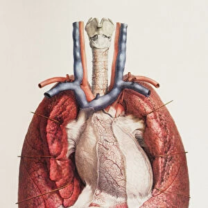
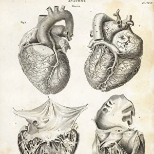
Heart. Historical anatomical artwork of the human heart, seen from the front

![]()
Wall Art and Photo Gifts from Science Photo Library
Heart. Historical anatomical artwork of the human heart, seen from the front
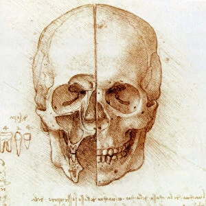
Heart. Historical anatomical artwork of the human heart, seen from the front. Coronary blood vessels are seen on the surface of the heart, supplying this muscular organ with oxygen so it can pump blood around the body. Blood vessels with deoxygenated blood are blue, while those with oxygenated blood are red. The blood vessels across top are (from left): the vena cava (blue), the aortic arch (red), and the pulmonary artery (blue). The pulmonary vein (not clearly seen) is to the right and behind the pulmonary artery. Veins bring blood to the heart. Arteries carry blood away from the heart. The pulmonary blood vessels carry blood between the heart and lungs. Artwork from Atlas of Anatomy, by Bourgery and Jacob, published in France in 8 volumes from 1831 to 1854
Science Photo Library features Science and Medical images including photos and illustrations
Media ID 6419534
© MEHAU KULYK/SCIENCE PHOTO LIBRARY
Anatomical Artwork Anterior Arteries Blood Circulation Blood Vessels Cardiac Cardiology Chest Coronary Dissected Dissection Drawing French Frontal Jacob Jean Baptiste Marc Bourgery Nicolas Henri Jacob Thoracic Thorax Veins Artery Circulatory System Courage Vein
FEATURES IN THESE COLLECTIONS
> Arts
> Artists
> J
> Jacob Jacobs
> Science Photo Library
> History
EDITORS COMMENTS
This print showcases a historical anatomical artwork of the human heart, providing us with a glimpse into the intricate beauty and complexity of our most vital organ. From the front view, we can observe the coronary blood vessels gracefully traversing the heart's surface, ensuring its oxygen supply for efficient blood pumping throughout our bodies. The artist's meticulous attention to detail is evident in their portrayal of deoxygenated blood vessels as serene blue pathways and oxygenated ones as vibrant red channels. Positioned across the top are significant blood vessels: the vena cava in blue, symbolizing veins bringing blood to the heart; followed by the aortic arch in red, representing arteries carrying life-sustaining oxygen away from this courageous organ; finally culminating with another blue vessel known as the pulmonary artery responsible for transporting blood between our hearts and lungs. Dating back to 19th-century France, this remarkable illustration originates from Jean Baptiste Marc Bourgery and Nicolas Henri Jacob's renowned "Atlas of Anatomy". Their comprehensive work spanning eight volumes offers invaluable insights into medical history and biological understanding. As we delve into this mesmerizing image capturing both artistry and scientific precision, we gain a deeper appreciation for how our circulatory system functions harmoniously within our thoracic cavity. This single snapshot encapsulates centuries of knowledge passed down through generations—a testament to humanity's unyielding pursuit of unraveling nature's mysteries.
MADE IN THE USA
Safe Shipping with 30 Day Money Back Guarantee
FREE PERSONALISATION*
We are proud to offer a range of customisation features including Personalised Captions, Color Filters and Picture Zoom Tools
SECURE PAYMENTS
We happily accept a wide range of payment options so you can pay for the things you need in the way that is most convenient for you
* Options may vary by product and licensing agreement. Zoomed Pictures can be adjusted in the Cart.