Home > Popular Themes > DNA
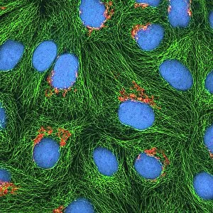
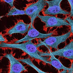
Mitosis, light micrograph

![]()
Wall Art and Photo Gifts from Science Photo Library
Mitosis, light micrograph
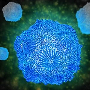
Mitosis. Confocal light micrograph of the stages of mitosis (nuclear division) and cytokinesis (cell division). During mitosis the nuclear envelope disintegrates (3rd image) and the chromosomes (blue), which are made of deoxyribonucleic acid (DNA), line up along the centre of the cell (4th image). Microtubules (red) attach to the chromosomes and pull them apart to opposite poles of the cell (5th image). The nucleus reforms and the cell begins to divide into two (last two images). This process is completed during cytokinesis to form two daughter cells. Magnification: x500 when printed at 10 centimetres wide
Science Photo Library features Science and Medical images including photos and illustrations
Media ID 6454595
© THOMAS DEERINCK, NCMIR/SCIENCE PHOTO LIBRARY
Anaphase Cell Division Cellular Chromatid Chromatids Chromosome Chromosomes Confocal Micrograph Cycle Cytokinesis Cytological Cytology Cytoplasmic Cleavage Cytoskeletal Daughter Dividing Division Furrow Hela Cell Immunofluorescence Immunofluorescent Interphase Metaphase Microtubules Mitosis Mitotic Nuclear Nuclei Nucleus Plasma Membrane Prometaphase Prophase Segregating Segregation Separating Stage Stages Telophase Deoxyribonucleic Acid Genetics Light Micrograph Light Microscope
FEATURES IN THESE COLLECTIONS
> Popular Themes
> DNA
EDITORS COMMENTS
This print showcases the intricate process of mitosis, captured through a confocal light micrograph. The image depicts the various stages of nuclear division and cell division, known as cytokinesis. As the third image reveals, the nuclear envelope disintegrates while in the fourth image, chromosomes (blue) composed of DNA align along the center of the cell. Fascinatingly, microtubules (red) can be observed attaching to these chromosomes in the fifth image and pulling them apart towards opposite poles of the cell. Gradually, as shown in subsequent images, the nucleus reforms and initiates cell division into two daughter cells. With a magnification level of x500 when printed at 10 centimeters wide, this photograph offers an up-close view into this crucial biological phenomenon. It highlights how cells undergo segregation and separation during mitosis while emphasizing key elements such as nuclear furrow formation and chromosome movement. The significance of this process extends beyond mere cellular replication; it plays a vital role in growth, development, repair, and reproduction within living organisms. Through this stunning visual representation captured by Science Photo Library's expertise in cytology and immunofluorescent techniques with light microscopy technology—this print serves as a testament to their commitment to scientific exploration.
MADE IN THE USA
Safe Shipping with 30 Day Money Back Guarantee
FREE PERSONALISATION*
We are proud to offer a range of customisation features including Personalised Captions, Color Filters and Picture Zoom Tools
SECURE PAYMENTS
We happily accept a wide range of payment options so you can pay for the things you need in the way that is most convenient for you
* Options may vary by product and licensing agreement. Zoomed Pictures can be adjusted in the Cart.