Home > Arts > Artists > G > Thomas Gray
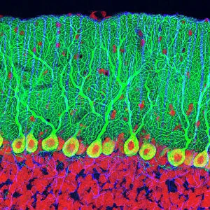
Purkinje nerve cells in the cerebellum

![]()
Wall Art and Photo Gifts from Science Photo Library
Purkinje nerve cells in the cerebellum
Purkinje cells in the cerebellum. Fluorescent light micrograph of Purkinje cells (green) in the cerebellum of the brain. Purkinje nerve cells have a flask-like body from which numerous highly branched dendrites extend. They are found in the grey matter (cortex) of the cerebellum, at the boundary between the granular layer (blue/red) and the molecular layer (red/green). The dendrites relay signals to the cell body, which passes them on through its single axon (green) in the granular layer. The cerebellum is a structure at the base of the brain that plays an important role in motor control, sensory perception and learning. Magnification: x200 when printed 10cm wide
Science Photo Library features Science and Medical images including photos and illustrations
Media ID 6449277
© THOMAS DEERINCK, NCMIR/SCIENCE PHOTO LIBRARY
Cerebellar Cerebellum Cortex Dendrite Dendrites Fluorescent Light Micrograph Granular Layer Gray Grey Matter Histological Histology Molecular Layer Nerve Cell Neuron Purkinje Cell Brain Light Microscope Nervous System Neurological Neurology
FEATURES IN THESE COLLECTIONS
> Animals
> Mammals
> Muridae
> Blue-grey Mouse
> Arts
> Artists
> G
> Thomas Gray
> Science Photo Library
> Specialist Imaging
EDITORS COMMENTS
This image showcases a fluorescent light micrograph of Purkinje cells in the cerebellum of the human brain. Purkinje cells, named after the Czech anatomist Jan Evangelista Purkinje who first described them in 1837, are large, flask-shaped nerve cells located in the grey matter (cortex) of the cerebellum. They are easily distinguishable due to their unique morphology, characterized by a large, flask-like soma from which numerous highly branched dendrites extend. The dendrites of Purkinje cells are the primary site of reception and integration of signals from the granular layer below, which are then relayed to the cell body. The cell body passes these signals on through its single axon, which extends into the granular layer and forms synapses with the dendrites of granule cells. The cerebellum, a structure located at the base of the brain, plays a crucial role in motor control, sensory perception, and learning. In this image, the Purkinje cells are situated at the boundary between the granular layer (stained blue/red) and the molecular layer (stained red/green). The molecular layer is where the Purkinje cell axons form synapses with mossy fibers, initiating the complex neural networks that underpin the functions of the cerebellum. Magnified at 200x when printed 10cm wide, this mesmerizing snapshot offers a glimpse into the intricate world of the human nervous system. The delicate balance and intricate connections between these cells underscore the complexity and sophistication of the brain's neural architecture.
MADE IN THE USA
Safe Shipping with 30 Day Money Back Guarantee
FREE PERSONALISATION*
We are proud to offer a range of customisation features including Personalised Captions, Color Filters and Picture Zoom Tools
SECURE PAYMENTS
We happily accept a wide range of payment options so you can pay for the things you need in the way that is most convenient for you
* Options may vary by product and licensing agreement. Zoomed Pictures can be adjusted in the Cart.