Inner ear anatomy C018 / 6379

![]()
Wall Art and Photo Gifts from Science Photo Library
Inner ear anatomy C018 / 6379
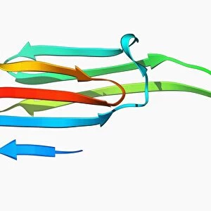
Inner ear anatomy. Computer artwork of a section through the inner part of a human ear, the organ of hearing and balance. The eardrum transmits sounds, as vibrations, from the air to the bones (ossicles), of the middle ear. These bones, from left to right, are the malleus (hammer), incus (anvil) and stapes (stirrup). The ossicles join to the inner ear, which consists of fluid-filled passages called the labyrinth (blue). This includes the cochlea (spiral), which translates the vibrations into electrical impulses that are carried to the brain by nerves (orange), and the semi-circular canals, which are responsible for balance
Science Photo Library features Science and Medical images including photos and illustrations
Media ID 9235933
© PASIEKA/SCIENCE PHOTO LIBRARY
Auditory Nerve Auditory Sense Aural Balance Cochlea Cochlear Ear Canal Eardrum Hammer Hearing Incus Labyrinth Malleus Nerve Nerves Ossicle Ossicles Stapes Stirrup Tympanic Membrane
EDITORS COMMENTS
This print showcases the intricate inner ear anatomy, a vital organ responsible for both hearing and balance. In this computer artwork, we are granted a glimpse into the hidden world of our auditory system. The eardrum takes center stage as it dutifully transmits sound vibrations from the air to the three tiny bones of the middle ear: the malleus (hammer), incus (anvil), and stapes (stirrup). These delicate ossicles form a crucial link between the outer and inner ear. The inner ear, depicted in mesmerizing shades of blue, is composed of fluid-filled passages known as the labyrinth. Within this labyrinth lies an extraordinary structure called the cochlea, resembling a spiral staircase. It is here that vibrations are transformed into electrical impulses by specialized cells, allowing us to perceive sound. Accompanying these remarkable structures are vibrant orange nerves that carry these electrical signals to our brain for interpretation. Additionally, we catch sight of another component essential for maintaining equilibrium -the semi-circular canals- which play a pivotal role in our sense of balance. Through this awe-inspiring image captured by PASIEKA/SCIENCE PHOTO LIBRARY, we gain insight into how our ears function on an anatomical level. It serves as a reminder that within each individual lies an intricately designed auditory system capable of perceiving sounds with astounding precision and maintaining harmony within ourselves.
MADE IN THE USA
Safe Shipping with 30 Day Money Back Guarantee
FREE PERSONALISATION*
We are proud to offer a range of customisation features including Personalised Captions, Color Filters and Picture Zoom Tools
SECURE PAYMENTS
We happily accept a wide range of payment options so you can pay for the things you need in the way that is most convenient for you
* Options may vary by product and licensing agreement. Zoomed Pictures can be adjusted in the Cart.