Human arteries, 19th Century illustration

![]()
Wall Art and Photo Gifts from Science Photo Library
Human arteries, 19th Century illustration
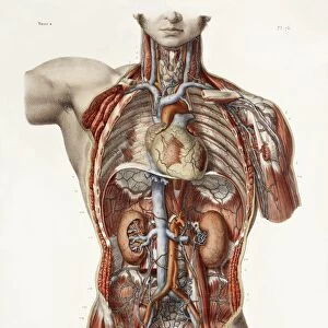
Human arteries, 19th Century illustration. Historical hand coloured lithographic print showing the arteries (red) and muscle structure (brown) of the human torso. The aorta (red) can be seen running down from the aortic arch (A, upper centre) as the descending aorta (B) and into the abdominal cavity (abdominal aorta, C), at the bottom of which it divides at the aortic bifurcation into the iliac arteries (lower left and right). Image from Traite complet de l anatomie de l homme, comprenant la medecine operatoire Vol. 4 (1836), by Jean-Baptiste Marc Bourgery and illustrated by Nicolas-Henri Jacob
Science Photo Library features Science and Medical images including photos and illustrations
Media ID 6327929
© SCIENCE PHOTO LIBRARY
1836 Abdomen Abdominal Arteries Axillary Body Carotid Chest Circulatory Descriptive Anatomy Diagram French Frontal Iliac Inferior Intercostal Internal Lithograph Lithographic Print Mammary Muscle Structure Muscles Plate 14 Shoulder Shoulders Superficial Thoracic Thorax Torso Transverse Trunk Vascular System Vessels Vol 4 Volume Four Wall Artery Circulation Mesenteric Artery Musculature Pelvis Subclavian
EDITORS COMMENTS
This 19th-century illustration offers a fascinating glimpse into the intricate network of human arteries. With its historical charm and meticulous detail, this hand-colored lithographic print showcases the internal structure of the torso with remarkable accuracy. The vibrant red hue highlights the path of the arteries, while shades of brown depict the underlying muscle structure. At the center of attention is the aorta, prominently running down from the aortic arch in an elegant curve. As it descends into the abdominal cavity, it divides at the aortic bifurcation to form two iliac arteries on either side. This comprehensive diagram also reveals other significant vessels such as carotid, subclavian, mesenteric artery, and mammary arteries. The image originates from "Traite complet de l'anatomie de l'homme" authored by Jean-Baptiste Marc Bourgery and illustrated by Nicolas-Henri Jacob in 1836. It serves as an invaluable resource for descriptive anatomy during that era. Displayed against a clean white background, this lithographic print exudes both scientific precision and artistic beauty. Its educational value lies not only in understanding our circulatory system but also appreciating how medical knowledge has evolved over time. Whether you are fascinated by biology or simply appreciate historical artwork, this print invites you to explore and marvel at one of nature's most incredible creations – our own bodies.
MADE IN THE USA
Safe Shipping with 30 Day Money Back Guarantee
FREE PERSONALISATION*
We are proud to offer a range of customisation features including Personalised Captions, Color Filters and Picture Zoom Tools
SECURE PAYMENTS
We happily accept a wide range of payment options so you can pay for the things you need in the way that is most convenient for you
* Options may vary by product and licensing agreement. Zoomed Pictures can be adjusted in the Cart.