Home > Animals > Mammals > Muridae > Water Mouse
Brain fibres, DTI MRI scan C017 / 7099

![]()
Wall Art and Photo Gifts from Science Photo Library
Brain fibres, DTI MRI scan C017 / 7099
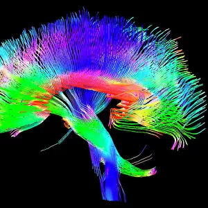
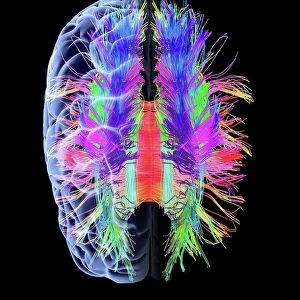
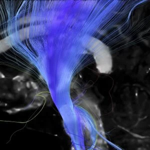
Brain fibres. 3D diffusion tensor imaging (DTI) magnetic resonance imaging (MRI) scan of nerve pathways in the brain. The pathways are highlighted in green and blue. The brain is seen from the front. Diffusion tensor imaging measures the direction of water diffusion, which in the brain reveals the orientation of nerve fibres. The technique is also known as tractography, with the resulting image known as a tractogram
Science Photo Library features Science and Medical images including photos and illustrations
Media ID 9340831
© SHERBROOKE CONNECTIVITY IMAGING LAB/SCIENCE PHOTO LIBRARY
Brain Imaging Brain Scan Central Nervous System Cerebral Cerebrum Diffusion Tensor Imaging Dti Scan Fiber Fibers Fibre Fibres Imaging Technique Magnetic Resonance Imaging Mri Scan Mri Scanner Nerve Nerve Fibre Nerves Neural Pathway Neural Tract Paths Pathway Pathways Structural Tractogram Tractography White Matter Brain Neurological Neurology
FEATURES IN THESE COLLECTIONS
> Animals
> Mammals
> Muridae
> Water Mouse
> Posters
> Scientific Posters
EDITORS COMMENTS
This print showcases the intricate network of brain fibres, as revealed by a 3D diffusion tensor imaging (DTI) magnetic resonance imaging (MRI) scan. The vibrant colours of green and blue highlight the pathways through which nerve signals travel within the brain. With a black background, this image exudes an air of mystery and fascination. Using the technique known as tractography, this MRI scan measures the direction of water diffusion in order to map out the orientation of nerve fibres. This innovative imaging technique provides valuable insights into the structural connectivity of our central nervous system. The complexity and beauty displayed in this image serve as a reminder of how remarkable our biological makeup truly is. It represents a visual representation of our neural pathways, illustrating how information is transmitted throughout our brain. With its focus on anatomy, medicine, and neurology, this print holds great significance for researchers and medical professionals alike. It offers a glimpse into normal brain function while also providing insight into potential abnormalities or disorders that may affect these vital pathways. Overall, this stunning photograph from Sherbrooke Connectivity Imaging Lab/Science Photo Library serves as both an artistic masterpiece and an invaluable scientific tool for understanding the complexities of human cognition and neurological health.
MADE IN THE USA
Safe Shipping with 30 Day Money Back Guarantee
FREE PERSONALISATION*
We are proud to offer a range of customisation features including Personalised Captions, Color Filters and Picture Zoom Tools
SECURE PAYMENTS
We happily accept a wide range of payment options so you can pay for the things you need in the way that is most convenient for you
* Options may vary by product and licensing agreement. Zoomed Pictures can be adjusted in the Cart.