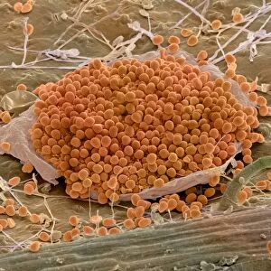
Athletes foot fungus, SEM

![]()
Wall Art and Photo Gifts from Science Photo Library
Athletes foot fungus, SEM
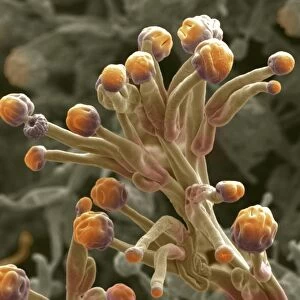
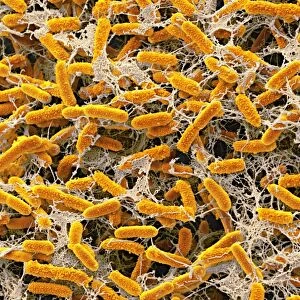
Athletes foot. Coloured scanning electron micrograph (SEM) of spores (yellow) of the fungus that causes athletes foot (tinea pedis) on skin (purple) from a human foot. The fungus grows on the surface of the skin and then into the living skin tissue itself, causing infection. It usually occurs between the toes, but may spread to the bottom and sides of the foot. The spores can lie dormant for long periods, until conditions are favourable for germination. Treatment involves keeping the infected area dry and using an anti-fungal powder. Magnification: x650 when printed centimetres wide
Science Photo Library features Science and Medical images including photos and illustrations
Media ID 6278265
© STEVE GSCHMEISSNER/SCIENCE PHOTO LIBRARY
Athletes Foot Dermatological Dermatology Dermatophyte Foot Fungal Fungi Fungus Infected Infection Keratin Micro Organism Microbe Mycology Re Production Reproductive Skin Spore Spores Abnormal Condition Disorder Health Care Micro Biology Microbiological Tinea Pedis Trichophyton Unhealthy
EDITORS COMMENTS
This print showcases the microscopic world of athletes foot fungus, offering a vivid insight into the nature of this common condition. The coloured scanning electron micrograph (SEM) reveals yellow spores belonging to the fungus responsible for athletes foot, known as tinea pedis. These spores can be seen scattered across purple skin tissue from a human foot. Athletes foot typically thrives on the surface of the skin before infiltrating deeper layers, causing infection and discomfort. Although it commonly occurs between toes, it can spread to other areas such as the bottom and sides of the foot. Interestingly, these resilient spores have an ability to remain dormant for extended periods until favorable conditions prompt their germination. The treatment for athletes foot involves maintaining dryness in the infected area and utilizing anti-fungal powder. This image serves as a reminder that even seemingly insignificant organisms like fungi can disrupt our well-being if given an opportunity. With a magnification level of x650 when printed centimeters wide, this photograph provides valuable insights into both biology and dermatology. It highlights how understanding microorganisms is crucial in healthcare practices while shedding light on various aspects such as reproduction, infection, and abnormality within our own bodies. Captured by Science Photo Library using a scanning electron microscope (SEM), this visually striking image combines scientific precision with artistic flair to create an intriguing visual representation of this common yet bothersome condition affecting many individuals worldwide.
MADE IN THE USA
Safe Shipping with 30 Day Money Back Guarantee
FREE PERSONALISATION*
We are proud to offer a range of customisation features including Personalised Captions, Color Filters and Picture Zoom Tools
SECURE PAYMENTS
We happily accept a wide range of payment options so you can pay for the things you need in the way that is most convenient for you
* Options may vary by product and licensing agreement. Zoomed Pictures can be adjusted in the Cart.